






 Portuguese


Device Developed by CNPEM Cultivates 3D Cells, Allows Flow Testing, and Recovers Intact Models for Further Analysis.
A microscopic device developed by researchers at the National Center for Energy and Materials Research (CNPEM) enables the cultivation of three-dimensional cell models, allows exposure to various substances, and permits the extraction of these structures from the platform after experiments without damaging them. This technology was designed to make toxicity studies more representative of living organisms and to expand alternatives to animal testing in research.
The work was published in July in the scientific journal ACS Measurement Science Au and is part of research from the Molecular Engineering Research Center for Advanced Materials (CEMol), based at CNPEM and funded by FAPESP as a Research, Innovation, and Dissemination Center. The platform will potentially be able to support studies involving pharmaceuticals, new materials, and environmental contaminants.
Device Can Be Opened After the Experiment Ends
One of the key features that differentiates this technology is its ability to disassemble the platform at the end of an experiment. In many microscopic systems, recovering a cell structure cultivated within the channels can be challenging, limiting the types of analyses that can be performed after exposure to a particular substance.
-
CERN Scientists “See” Structures Smaller Than a Proton and Discover Signs of a Phenomenon Predicted by Physics

-
236-Million-Year-Old Fossil May Change Mammal History by Revealing Dog-Sized Ancestor Gave Birth to Live Young
-
1.27 Billion-Year-Old Martian Meteorite Fills Nearly 2 Billion-Year Gap in Mars’ History
-
One of the Most Difficult Plastics to Recycle May Gain New Life


In the device created at CNPEM, three-dimensional models can be extracted intact for a subsequent investigation phase. This enables, for example, a more detailed observation of the changes that remain in the cells after contact with a compound, rather than restricting research to data obtained while the experiment is still ongoing.
In a statement to CNPEM’s Press Office, Iris Renata Sousa Ribeiro, a postdoctoral researcher at the National Nanotechnology Laboratory (LNNano-CNPEM) and the study’s lead author, emphasized this expansion of analytical possibilities after the experiment.
Microscopic Flow Attempts to Recreate Conditions Cells Encounter in the Body
The platform employs microfluidics, a field that utilizes very small channels to control the movement of liquids. In this environment, researchers can precisely determine how nutrients, oxygen, and experimental substances reach the cells throughout the test.
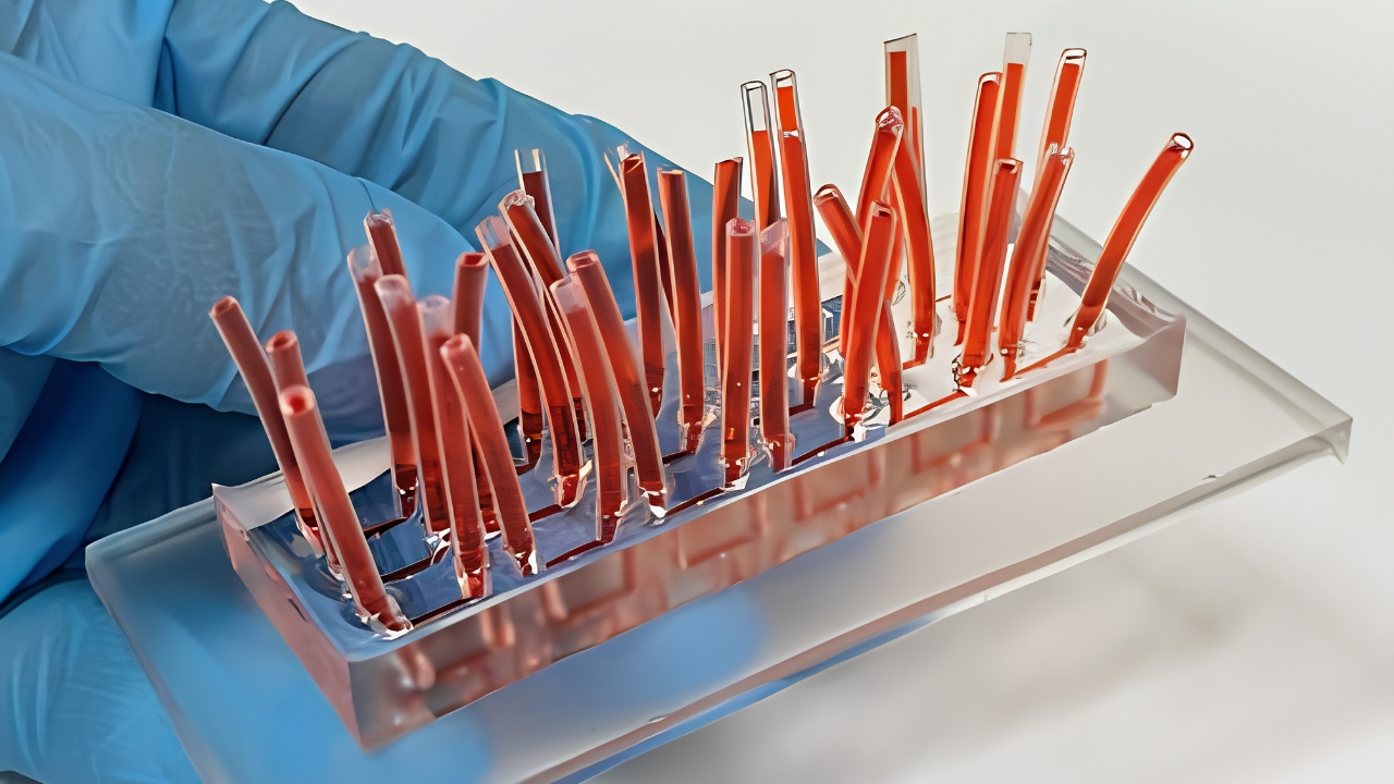
The device maintains this movement continuously, creating a condition different from that found in static cell cultures. In living tissues, molecules and nutrients are constantly being transported throughout the organism; replicating part of this dynamic in the lab can generate responses that are closer to actual biological conditions.
This aspect is especially important for toxicity studies. The way a substance reaches the cells, remains available, and interacts with the tissue can alter the observed effects during an experiment.
3D Models Offer Different Insights Beyond Flat Cultures
The technique also alters the organization of the studied cells. Traditional laboratory cultures typically grow on two-dimensional surfaces, while the models used in the new platform form three-dimensional structures known as spheroids.
In these structures, the cells establish contacts in multiple directions, a condition much closer to the architecture found in tissues. Therefore, the device can be employed to investigate responses to drugs, nanomaterials, and potentially harmful agents to the environment without relying exclusively on flat cultures.
The platform is made from PDMS, a transparent and biocompatible silicone. The material is also flexible and relatively low-cost, characteristics that favor the production of microfluidic systems intended for repeated experiments.
Technology Designed for Laboratories without Microfluidics Specialists
Another goal of the researchers was to reduce the technical barrier for using the system. The team established a standardized experimental protocol so that groups not routinely working with microfluidics could incorporate the device into cellular assays.
According to Ribeiro, the intention was to create a simple and reproducible procedure that could be adopted by professionals in various fields. This standardization is important because laboratory technologies that heavily depend on specialists may remain restricted to the groups that directly participated in their development.
The team is working to transform the platform into infrastructure available to external researchers by early next year. Universities, research institutes, and companies are among the audiences that will be able to utilize the resource.
Device Could Support Research with Highly Diverse Goals
The opening of the infrastructure should allow the same system to be applied to distinct scientific questions. Instead of functioning solely as a platform aimed at one type of test, the project was designed to accommodate studies in multiple areas.
Among the anticipated uses are:
- ecotoxicology, assessing how pollutants and other substances may affect cellular systems;
- pharmacology, investigating responses and toxicity of drug candidates;
- materials science, examining the interaction between new materials and cells;
- nanotechnology, monitoring the biological effects produced by nanoscale structures;
- biotechnology, developing cellular assays that depend on controlled flow conditions.
The list demonstrates how the same infrastructure can address different questions without altering its central principle: to keep cells organized in three dimensions while controlling the surrounding environment at a microscopic scale.
Recovering Cells Can Reveal What Happened After Exposure
The scientific gain does not end when the flow is interrupted. By removing the intact spheroids, researchers can subject them to complementary techniques and investigate mechanisms that would not be visible solely during the initial exposure.
This design enhances the experimental value of each test and can help understand not only if a substance produces toxic effects but also how this alteration develops within the cellular model.
The study received support from FAPESP through different research projects and is part of the effort by the Molecular Engineering Research Center for Advanced Materials (CEMol) to develop tools aimed at advanced materials and biological systems. With the future opening of the platform to external users, the National Center for Energy and Materials Research (CNPEM) aims to take the device beyond the laboratory where it was developed and transform it into a shared tool for more controlled, reproducible tests that closely mimic conditions found in living organisms.


