 Portuguese
  English
  Spanish
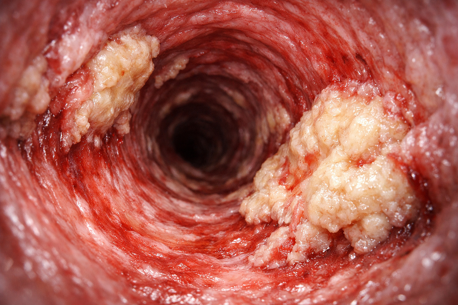
Unpublished Images Captured Inside Arteries Show, In Detail, How PCSK9 Inhibitor Drugs Manage to Reduce, Stabilize Plaques and Explain the Real Drop in the Risk of Heart Attacks and Strokes
For decades, cardiologists had to interpret indirect signals to understand what was happening inside the heart’s arteries. Blood tests indicated cholesterol levels, while angiograms only showed how narrowed the vessel was. However, nothing accurately revealed what was hidden in the arterial wall. Now, this scenario is changing. With the use of microcameras inserted directly into the coronary arteries, doctors can observe, almost in real time, how modern medications act directly on dangerous atherosclerotic plaques.
The information was disclosed by Current Atherosclerosis Reports, in a scientific review that gathered data from major international studies. As researchers demonstrated, the so-called PCSK9 inhibitors, when combined with traditional statins, not only reduce LDL cholesterol in the blood but also promote visible structural changes in the plaques within the arteries over approximately one year of treatment.
These findings help clarify why patients using these drugs experience fewer heart attacks and strokes, going beyond laboratory numbers and showing physical evidence of stabilization of coronary artery disease.
-
A gigantic project in the Netherlands is using the sediment from a degraded lake to create artificial islands, improve water quality, and restore natural shorelines.

-
After seeing a 100% automated factory in Shanghai, the president of Honda exclaimed, “we don’t stand a chance”; China accelerates electric cars, cuts costs, and exposes the brand’s crisis worldwide.

-
A giant half-ton ring fell in Kenya from space, and more than a year later, scientists still do not know where it came from.

-
Dubai eliminates the term “housewife” from official documents and adopts “generation builder,” redefining the role of mothers and changing the way society views motherhood.

What Doctors Can Finally See Inside the Heart’s Arteries
For many years, the main exam to assess coronary arteries was angiography. This method uses contrast to show only the internal space of the vessel, revealing where there is narrowing, but without detailing the contents of the arterial wall. Thus, dangerous plaques could go unnoticed, especially those that were not yet causing significant obstruction.
With technological advancements, recent studies have started to employ invasive examinations, in which a thin catheter with sensors is inserted directly into the artery. This has made it possible to precisely monitor the same arterial segment before and after treatment. This allowed researchers to assess whether the plaques were diminishing, becoming more stable, or continuing to evolve silently.
Major hospitals in Europe and North America gathered data from hundreds of patients. All had been using powerful statins and subsequently received a PCSK9 inhibitor. Throughout the follow-up, researchers observed consistent changes in the composition and volume of the plaques, something never documented so clearly until now.
How PCSK9 Inhibitors Act on Cholesterol and Plaques
LDL cholesterol, popularly known as “bad cholesterol,” circulates in the bloodstream and can deposit itself in the walls of the arteries. Over time, a plaque composed of fat, cholesterol crystals, and scar tissue forms. When this plaque grows or ruptures, blood flow can be blocked, resulting in a heart attack or stroke.
Statins remain the cornerstone of treatment, as they reduce LDL and significantly decrease cardiovascular risk. Nevertheless, many high-risk patients remain vulnerable, even with apparently controlled levels. It is in this context that PCSK9 inhibitors come in, helping the liver remove even more LDL from circulation.
In the clinical study FOURIER, for example, the addition of the medication evolocumab to statins reduced the average LDL to about 30 mg/dL. Moreover, there was a significant drop in severe cardiovascular events. From this result, a fundamental question arose: did the benefit come solely from improved numbers, or was there a real change inside the arteries?
Microcameras, Ultrasound, and Light Reveal the Transformation of Plaques
To answer this question, researchers turned to intravascular imaging technologies. The intravascular ultrasound (IVUS) uses sound waves to create cross-sectional images of the artery, allowing the measurement of how much space the plaque occupies in the arterial wall.
The optical coherence tomography (OCT) uses light to generate extremely detailed images of the so-called “fibrous cap,” a thin layer that covers the fatty nucleus of the plaque. The thinner this cap, the greater the risk of rupture. Therefore, an increase in thickness serves as a direct indicator of greater stability.
Additionally, the near-infrared spectroscopy (NIRS) analyzes the absorption of light by the plaques, estimating the amount of lipids present. By combining these techniques, doctors can identify not only the size of the plaque but also its composition and risk level.
Clinical Trials Tracking the Change of Plaques Over Time
The study GLAGOV evaluated nearly 1,000 patients on statin therapy. After initial exams with ultrasound, participants received evolocumab or placebo and were re-evaluated about 18 months later. The results showed a significant reduction in plaque volume, and more than half of the patients demonstrated real regression of atherosclerosis.
In the trial HUYGENS, which included patients after a heart attack without ST elevation, serial exams revealed that evolocumab increased the thickness of the fibrous cap and reduced the lipid content of the plaques compared to placebo.
The study PACMAN-AMI, involving approximately 300 patients treated after a classic heart attack, analyzed arteries that had not caused the initial event. In just one year, the use of alirocumab in combination with high-intensity statins reduced the total plaque volume, decreased the fatty nucleus, and thickened the protective cap.
Meanwhile, the study ARCHITECT, involving over 100 patients with familial hypercholesterolemia, used coronary computed tomography. After about 18 months, a reduction in total plaque burden was observed, along with a change in composition, with less unstable tissue and more fibrous and calcified material.
Why More Stable Plaques Mean Fewer Heart Attacks in the Future
Cardiologists know that not every plaque causes immediate symptoms. Many of the most dangerous do not cause significant obstruction and thus go unnoticed in traditional exams. However, characteristics such as a large lipid nucleus, thin cap, intense inflammation, and fragile tissue drastically increase the risk of rupture.
In the analyzed studies, PCSK9 inhibitors moved the plaques away from this high-risk profile. They became smaller, more fibrous, with less loose cholesterol and thicker caps. In simple terms, unstable and fragile plaques began to behave more like solid and predictable structures.
These changes help explain why maintaining very low LDL levels over time translates into fewer cardiovascular events, directly connecting laboratory data with real images inside the arteries.
The Impact on Patients and the Future of Cardiac Treatment
These discoveries are changing the way coronary artery disease is understood. Today, it’s not just the degree of narrowing of the artery that matters, but also the type of plaque present. A light obstruction with unstable plaque can be more dangerous than a greater narrowing formed by denser and more stable tissue.
Researchers are now investigating whether less invasive exams, such as advanced CT scans, can identify patients with still dangerous plaques even when traditional exams appear normal. This may allow the use of PCSK9 inhibitors to be directed more accurately, considering not only cholesterol but the biology of each individual’s plaque.
Ongoing trials are assessing the earlier use of these drugs after heart attacks and the possibility of incorporating intravascular imaging into future guidelines. For now, the conclusion is clear: in the right patients, these medications not only improve blood test results but promote a true visible repair in the heart’s arteries.
The study “PCSK9 and Coronary Artery Plaque: New Opportunity or Red Herring?” was published in the journal Current Atherosclerosis Reports.


-

-

3 pessoas reagiram a isso.