
 Portuguese
  Spanish
European XFEL is the world’s largest X-ray laser, generating 27,000 flashes per second and observing atoms, molecules, and extreme materials.
Observing molecules in motion, tracking chemical reactions almost in real-time, and recording invisible structures at atomic scales seemed like science fiction until recently. Today, this is already happening at the European XFEL, one of the most advanced scientific infrastructures on the planet. Located between Hamburg and Schenefeld, in Germany, the European XFEL began operations for users in 2017 and has established itself as the world’s largest X-ray laser.
The facility combines gigantic dimensions, ultra-short pulses, and extreme brightness to investigate phenomena that conventional microscopes simply cannot capture.
What is the European XFEL and why it became the world’s largest X-ray laser
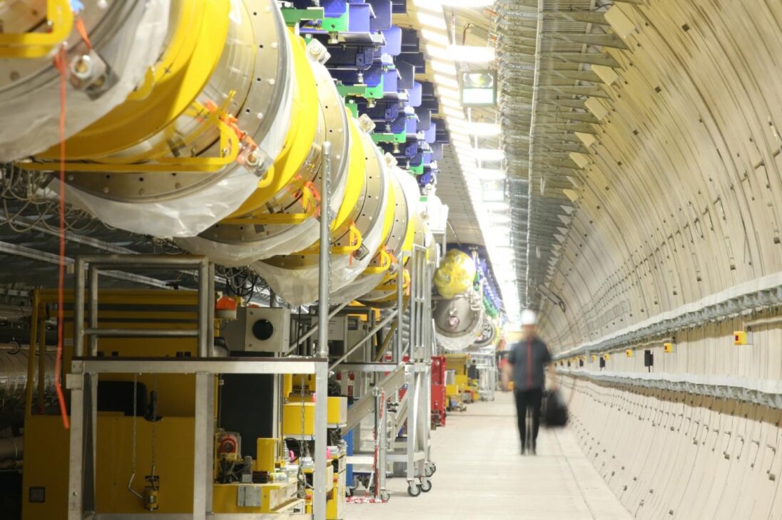
The European XFEL is an international scientific infrastructure installed mainly in underground tunnels.
-
Former SpaceX Rocket Engineer Develops Fast-Growing Grass Panels to Replace Wood in Construction, Secures $47.5 Million Funding

-
A Simple Finger Movement Gains Attention for Its Potential Role in Alzheimer’s Prevention

-
MSC Orders Largest Fleet of Gas-Powered Ships in Maritime History, Set to Impact Global Freight Costs

-
WhatsApp Introduces Two New Features to Revolutionize How Millions Add Contacts


According to DESY and the center itself, the facility is 3.4 kilometers long and connects the DESY campus in Hamburg to the municipality of Schenefeld, where the research campus and the large experimental area are located.
This physical scale is impressive, but size alone does not explain the importance of the machine. What makes the European XFEL unique is the ability to generate extremely intense and coherent X-rays to reveal details of matter at the atomic and molecular level.
In practice, it functions as a kind of ultra-fast camera of the nanoworld. Instead of observing only static structures, the system was designed to capture dynamic processes, including ultra-fast changes in molecules, materials, and biological systems.
How the European XFEL produces X-rays with extreme precision
The heart of the facility is a superconducting linear accelerator about 1.7 kilometers long, responsible for accelerating electrons to speeds very close to the speed of light.
After that, these particles pass through long undulators, magnetic structures that force the electrons to oscillate and emit X-ray pulses.

According to official data, the electrons in the system reach 99.99999996% of the speed of light. The wavelength produced ranges from 0.05 to 4.7 nanometers, a range that allows investigation of extremely small structures, well below what would be accessible with conventional optical techniques.
It is this combination of superconducting acceleration, magnetic control, and coherent radiation that allows the European XFEL to move from the field of indirect observation to the territory of ultrafast imaging on an atomic scale.
27 thousand flashes per second put the European XFEL on another level
One of the most impressive numbers of the facility is its repetition rate. The European XFEL generates 27 thousand X-ray flashes per second, a capability that conventional sources do not reach and which, according to DESY, offers a decisive advantage for many types of experiments.
The intensity of these pulses also deviates from the standard. Official sources report that the brightness can be about a billion times greater than that of the best conventional synchrotron radiation sources, which greatly enhances the sensitivity of measurements and the quality of the images obtained.
Furthermore, the pulses are ultrashort, reaching tens of femtoseconds. This time scale is crucial because it allows recording of very fast phenomena, such as molecular rearrangements, electronic transitions, and initial stages of chemical reactions that normally escape direct observation.
What is the European XFEL used for in chemistry, biology, and materials science
The scientific applications of the European XFEL go far beyond basic physics. According to the center itself, the beams can be used to film ultrafast processes, observe changes in the configuration of biomolecules, record atomic details of viruses, decipher the molecular composition of cells, and produce three-dimensional images of the nanoworld.
The infrastructure is also used to investigate materials subjected to extreme conditions, including processes similar to those occurring inside planets and stars.
This transforms the facility into a strategic tool for areas such as chemistry, structural biology, condensed matter physics, advanced materials, energy, and medicine.
Instead of just showing the final form of a molecule or a material, the European XFEL allows us to follow what happens during the transformation. That is why the idea of “filming the movement of atoms” has ceased to be an exaggerated metaphor and has become a real objective of several experiments conducted there.
Why the European XFEL is already one of the most important scientific machines of this century
The European XFEL was designed to serve research teams from various fields simultaneously. The facility brings together multiple experimental stations, specialized laboratories, and infrastructure aimed at studies that require extremely high precision, acquisition speed, and large data volumes.

Its impact is not only on the scale of engineering but in the change of scientific capability. The machine has opened access to research areas that were previously considered too difficult or simply inaccessible, especially when the challenge was to observe phenomena that were too fast or too small for traditional instruments.
Therefore, the European XFEL represents more than a technological record. It shows how the next frontier of science involves making visible what has remained hidden in the heart of matter for decades.


