
 Portuguese
  Spanish
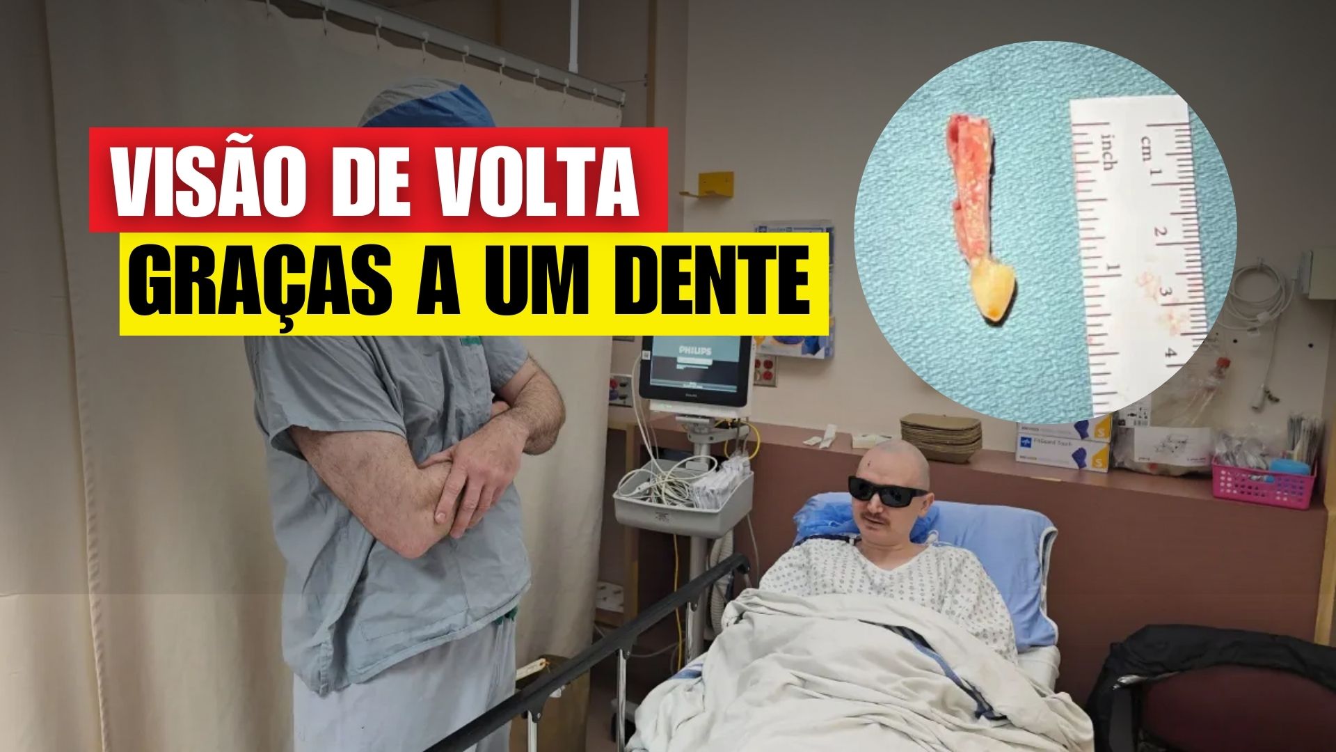
34-Year-Old Canadian Man Regained Sight After Two Decades Thanks to a Rare Technique Using a Tooth to Support a Lens in the Eye. The Procedure, Performed in Vancouver, Was the First Time the Surgery Took Place in Canada.
A 34-year-old Canadian man regained sight after two decades thanks to a rare technique using a tooth from the patient to support a lens in the eye.
The procedure, known as Osteo-Odonto-Keratoprosthesis (OOKP), was conducted in Vancouver by the team of ophthalmologist Greg Moloney from the University of British Columbia, and is part of the first series of surgeries of this kind performed in Canada.
According to information from CNN, with glasses, the patient achieved acuity of 20/30 in the right eye.
-
US Mayor Goes Undercover as Homeless for a Week to Understand Rising Homelessness, Sparking Debate in His City

-
Teen from Brazil Turns School Snack Sales into Million-Dollar Confectionery Business Projected to Earn Nearly $1.9 Million in 2024

-
Why Do Electrical Outlets and Switches Turn Yellow? Understanding the Impact of Light, Temperature, and Materials on Color Change
-
Former Brazilian laborer and ice cream vendor turns struggling company into $16.5 billion giant with 90,000 employees.


What Is the Tooth-in-the-Eye Technique
OOKP is indicated when corneal transplants fail and the ocular surface remains severely scarred, but the retina and optic nerve are preserved.
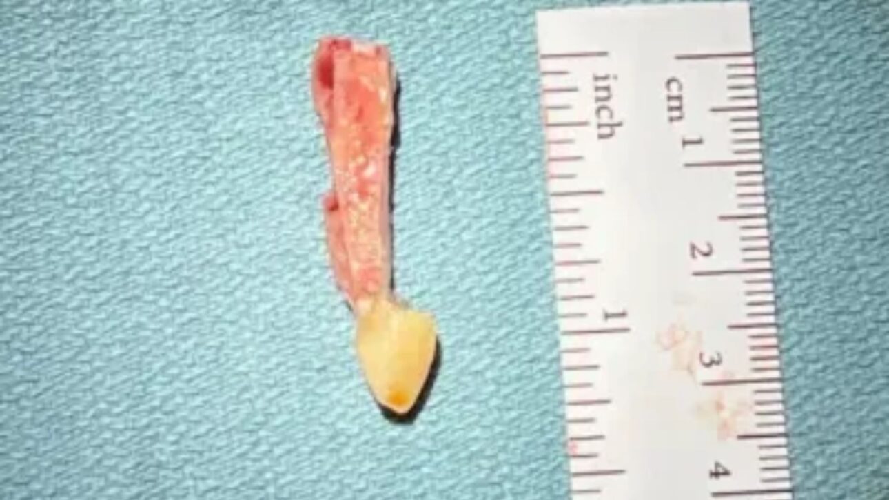
The method replaces the optical function of the damaged cornea with a structure made up of a tooth fragment and a small cylindrical lens, which fits into this graft.
As it is the patient’s own tissue, the structure has a higher chance of integration and a lower risk of rejection.
How the Tooth Becomes a Support for the Lens
First, surgeons extract a canine — usually with a thin layer of bone — and sculpt it into a block that receives, at the center, an optical cylinder.
Then, this structure is temporarily implanted in the cheek or eyelid for a few months to promote tissue growth and vascularization.
In the final stage, the tooth-lens block is fixed to the front part of the eye, opening up a pathway for light to reach the retina.
According to Moloney, “the tooth is an ideal structure to keep a focusing element in place: it is rigid, survives in adverse environments, and the body recognizes it as its own”.
Who Can Benefit
Cases of severe corneal blindness after chemical burns, trauma, or autoimmune diseases are among the main candidates.
In conditions like Stevens-Johnson Syndrome — triggered by infections or medications — limbal stem cells may be destroyed, leaving the cornea opaque and keratinized, which prevents light from passing.
In these scenarios, “it is a situation where a standard corneal transplant simply would not work”, explains ophthalmologist Vicente Diaz from Yale University.
The Patient’s Journey to Surgery
At 13 years old, during a Christmas basketball game, Brent Chapman suffered a severe drug reaction compatible with Stevens-Johnson Syndrome.
The episode caused skin burns and extensive damage to the ocular surface.
He lost the left eye due to infection and maintained residual vision in the right, which deteriorated over time.
In two decades, he tried dozens of procedures, mainly corneal transplants, without lasting results.
After evaluation by the Vancouver team, he was deemed suitable for OOKP: the “back” part of the eye was healthy, but the cornea could not tolerate new grafts.
The surgery was performed at Mount Saint Joseph Hospital in Vancouver, as part of the program that introduced the technique in the country.
Timeline of the Case
Treatment occurred in stages throughout 2025. In February, surgeons removed the canine and prepared the block with the lens.
In June, the structure was integrated into the right eye. On August 5, the team adjusted the alignment to correct distortions.
The patient received glasses on August 13 and began to see 20/30, a parameter indicating the ability to identify details at 6 meters that a person with perfect vision distinguishes at 9 meters.
The first clear image after the procedure was the horizon viewed from the 16th floor of the doctor’s office.
Rediscovering the World
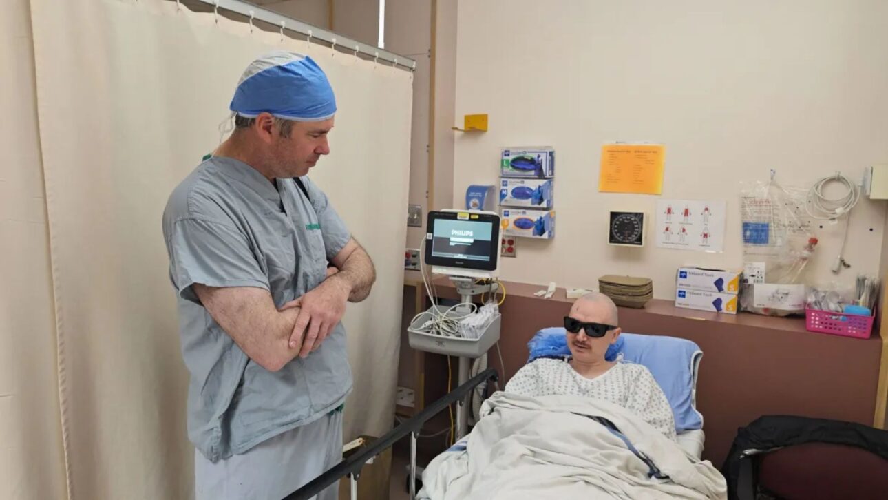
During immediate rehabilitation, Chapman reported feeling surprise and euphoria. “I am very happy and rediscovering the world, enjoying the little things,” he said.
When he saw the doctor after the final adjustment, he described a moment that marked him: “Dr. Moloney and I made eye contact for the first time, and we both got quite emotional”.
Why Use a Tooth
Besides mechanical resistance, the tooth offers structural stability for the lens and remains viable in environments with little lubrication — something common in eyes with severe scarring.
The team emphasizes that biocompatibility is a decisive factor for success, as the material belongs to the patient.
This strategy explains part of the good results in situations where artificial keratoprostheses or conventional grafts have failed.
A Rare and Time-Consuming Procedure
OOKP is a last-resort resource and requires specific training. Generally, the process is divided into two major surgeries, totaling over 12 hours, and involves integration between ophthalmology and buccal and maxillofacial surgery.
In Canada, the technique began to be offered this year by a small group of specialists. Patients who meet the criteria can regain near-normal vision.
The university responsible for the program confirms that the first cases in the country were completed in 2025, with varying levels of visual gain.
The Impact Outside the Surgical Center
With improved acuity, Chapman resumed daily tasks without constant support.
He started planning trips — with Japan on the list — and is preparing to return to work as a massage therapist.
The ability to make plans without fear of ocular emergencies was described by him as a psychological relief, after years of uncertainty and successive surgeries.
Nonetheless, it is important to highlight that specialists emphasize that success depends on appropriate selection: the retina and optic nerve need to be functional.


