Portuguese
  Spanish
Scientists create bioengineered esophagus that functions without rejection in an animal, advancement could transform human transplants.
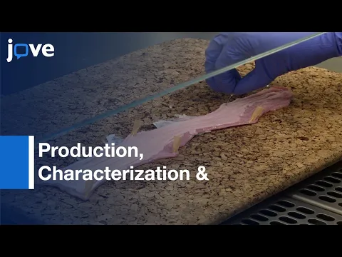
On March 20, 2026, researchers from Great Ormond Street Hospital in partnership with University College London announced a breakthrough that could redefine the future of transplants: the creation of a functional bioengineered esophagus, successfully implanted in a living animal, restoring swallowing without the need for immunosuppression. The information was officially released by the institution, which detailed the experiment as the most advanced ever documented in tubular organ engineering.
The study showed that the organ not only integrated into the animal’s body, but also grew with the organism and began to perform natural functions, something that until now was one of the main obstacles in regenerative medicine. The most relevant data is that the tissue was built with the recipient’s own cells, eliminating the risk of immunological rejection, which is currently one of the biggest limitations of traditional transplants.
Continue reading below to understand how this esophagus was created, why it functions without rejection, and how this advancement could directly impact the treatment of serious diseases in humans.
-
Brazilian Engineers Lead $200 Million Project to Bring Water from São Francisco River to Transform Supply in Northeastern Cities

-
Brazilian University Tests Recycled Plastic Component for Homes, Aiming for More Sustainable Single-Family Housing Construction
-
Apple, Samsung, and Sony Warn: Wet Bluetooth Earbuds Can Be Damaged if Stored Before Fully Drying
-
Brazil Develops AI-Driven Technology to Measure Brain Pressure Non-Invasively, Detecting Nanometric Deformations in Real Time to Aid Early Medical Intervention


Tissue engineering creates functional esophagus that replaces a complete part of the organ
The experiment carried out by British scientists achieved something medicine has been attempting for decades: replacing a complete section of a complex organ and making it function naturally.
The bioengineered esophagus was implanted in a growing animal, where it allowed the normal passage of food, restoring swallowing.
According to the released data, the organ was capable of:
- maintaining continuous food flow,
- integrating with surrounding tissues,
- developing functional structure over time.
This represents a significant technical leap, because tubular organs like the esophagus require mechanical coordination, elasticity, and integration with adjacent muscles and tissues.
Organ structure was created from a biological “scaffold” and the organism’s own cells
The process used did not simply involve “printing” an organ. Scientists used a technique known as decellularization and recellularization.
First, a biological esophagus structure of animal origin was used as a base, completely removing the original cells and retaining only the structural matrix.

Then, this structure was repopulated with cells from the recipient organism itself. This method allows the new organ to be recognized by the body as its own tissue. The result is an implant that drastically reduces the risk of rejection, one of the biggest challenges in transplant medicine.
Absence of immunosuppression marks a difference compared to traditional transplants
Today, transplant patients need to take immunosuppressive drugs for the rest of their lives. These medications prevent the body from attacking the transplanted organ but come with significant side effects, including an increased risk of infections and other complications.
In the case of the bioengineered esophagus, this was not necessary. Since the tissue was built with the organism’s own cells, the immune system did not recognize the organ as foreign.
This point is considered one of the most important advances of the study, as it paves the way for safer and more durable transplants.
Implanted organ grew with the animal and maintained functionality over time
Another historical challenge in organ engineering is ensuring that implanted tissue not only functions initially but continues to function over time.
In the experiment, the bioengineered esophagus:
- grew with the animal,
- maintained its structure,
- continued to allow normal feeding.
This behavior is essential, especially for pediatric applications. Children born with esophageal malformations need solutions that accompany body growth, something artificial prostheses cannot offer.
Esophageal atresia could be one of the first clinical applications of the technology
One of the main targets of this research is children with esophageal atresia, a congenital condition in which the esophagus does not form correctly.
In more severe cases, known as long-segment atresia, the organ lacks sufficient connection between its parts. Today, treatment involves complex surgeries that may use parts of the stomach or intestine to replace the esophagus.
These solutions, while functional, can lead to complications throughout life. The possibility of creating an esophagus with the patient’s own cells represents a potentially more efficient and less invasive alternative.
Engineering of hollow organs represents one of the greatest challenges in regenerative medicine
Creating solid organs, such as skin or cartilage, is already possible in some medical contexts. However, hollow organs, such as the esophagus, intestine, and trachea, present additional challenges.
They need to:
- maintain structural shape,
- allow passage of fluids or food,
- resist pressure and movement,
- integrate with adjacent tissues.
Therefore, the success of the experiment is considered a milestone. It demonstrates that it is possible to advance in the reconstruction of complex structures, which broadens the horizon of regenerative medicine.
Advancement is still in experimental phase and has not been applied in humans
Despite the promising results, it is important to highlight that the study was conducted in an animal model. To date, there has been no clinical application in humans.
Researchers indicate that the next step involves additional tests, safety validation, and the development of protocols for clinical use. This process could take years, depending on regulation and future results.
Data obtained can accelerate the development of other bioengineered organs
The knowledge generated by the study is not limited to the esophagus. The techniques used can be adapted for other tubular organs and structures of the human body. This includes future possibilities such as:
- tracheas,
- parts of the intestine,
- blood vessels.
The ability to create personalized tissues with the patient’s own cells can transform various areas of medicine.
Transplants may change model over the next decades
If the results are confirmed in humans, the impact could be structural. The current transplant model depends on donors, compatibility, and continuous use of medication.
With tissue engineering, the possibility arises of creating organs on demand. This could reduce transplant waiting lists, eliminate rejection, and increase the life expectancy of patients with serious diseases.
The study represents a paradigm shift. Instead of merely treating symptoms or repairing damaged structures, medicine is moving towards the complete replacement of organs.
This advancement is still in its early stages, but the results indicate that the direction is technically viable.
Given this scenario, the direct question that arises is: are we close to an era where human organs can be custom-produced in the laboratory, or are we still facing an advancement that will take decades to reach clinical practice?