
 Portuguese
  Spanish
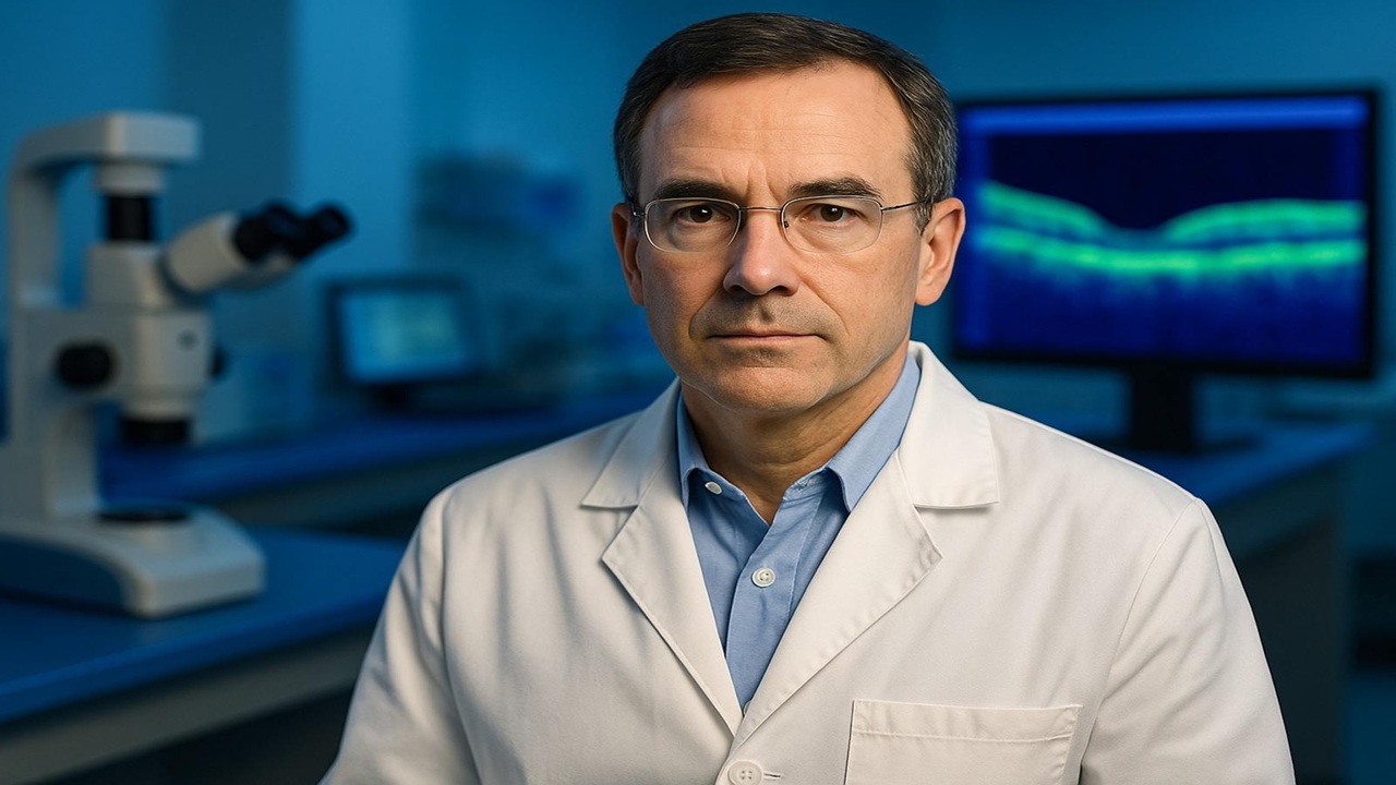
The First Human Trial with Retinal Pigment Epithelial Stem Cells Shows Full Safety and Significant Visual Improvement in Elderly with Advanced Dry AMD
Taking a photo and focusing on someone’s face or keeping one’s eyes on the road are actions that depend on central vision. For adults with age-related macular degeneration, this essential area of the retina loses sharpness to the point where details become unrecognizable.
The available alternatives only slow the progression or offer limited improvement, while blurriness tends to advance over time.
Early Trials Indicate Safety and Visual Advancement
A recent clinical trial evaluated a procedure based on stem cell transplantation with the potential to reverse accumulated damage to the macula, responsible for what is seen directly in front. This was the first application of this type of treatment in humans. As it is a phase 1/2a study, the investigation focused on safety and preliminary efficacy.
-
Helicopter Drops 180 Tons of Sand and Gravel on Swedish River to Revive Ecosystem Damaged by Decades of Exploitation

-
Brazilian Company Launches Retro Smartphone with 48 MP Camera, T9 Keyboard, and Privacy Features for Social Media-Free Use

-
Self-Taught Chinese Farmer Builds 5-Ton Submarine from Scrap, Launches It in Anhui River
-
Invisible Induction Technology Enables Wireless Power for Blenders, Coffee Makers, and Air Fryers, Reducing Countertop Cables


Researchers await broader comparisons with existing therapies, something expected only after phase 3. Nevertheless, initial results allowed the continuation of testing. Previous laboratory trials showed that transplanted cells preserved retinal identity and exhibited no signs of tumor or toxicity, which supported the decision to proceed.
After evaluating 18 potential candidates, the team selected six volunteers aged 71 to 86 diagnosed with the dry form of macular degeneration, responsible for about 80% of cases. Three of them had visual acuity between 20/200 and 20/800, while the others ranged from 20/70 to 20/200. The dry form presents progressive loss caused by small deposits of fats and proteins that damage retinal pigment epithelial cells.
Procedure and Improvement Registered Over Follow-Up
The method uses stem cells obtained from an eye bank, capable of producing new retinal pigment epithelial cells. Each participant received 50,000 cells through a single injection administered under the retina of the most affected superior temporal macula.
The trial recorded safety, with no evidence of tumors or immune reactions. Some typical complications of eye surgeries were observed, but no events related to the stem cells occurred. All volunteers exhibited improvement in vision in the treated eye, an improvement that was not identified in the other eye.
One year after the procedure, the three participants with the lowest initial acuity saw an average of 21 more letters on the Snellen chart. Rajesh Rao, physician-scientist and ophthalmologist at Michigan Medicine, stated that the gain exceeded expectations for advanced cases of the disease.
Next Steps of the Research
The study continues to monitor patients who received larger doses of 150,000 and 250,000 cells. If these amounts also prove safe, the team may expand testing in humans. The research was published in the journal Cell Stem Cell.


Be the first to react!