
 Portuguese
  Spanish
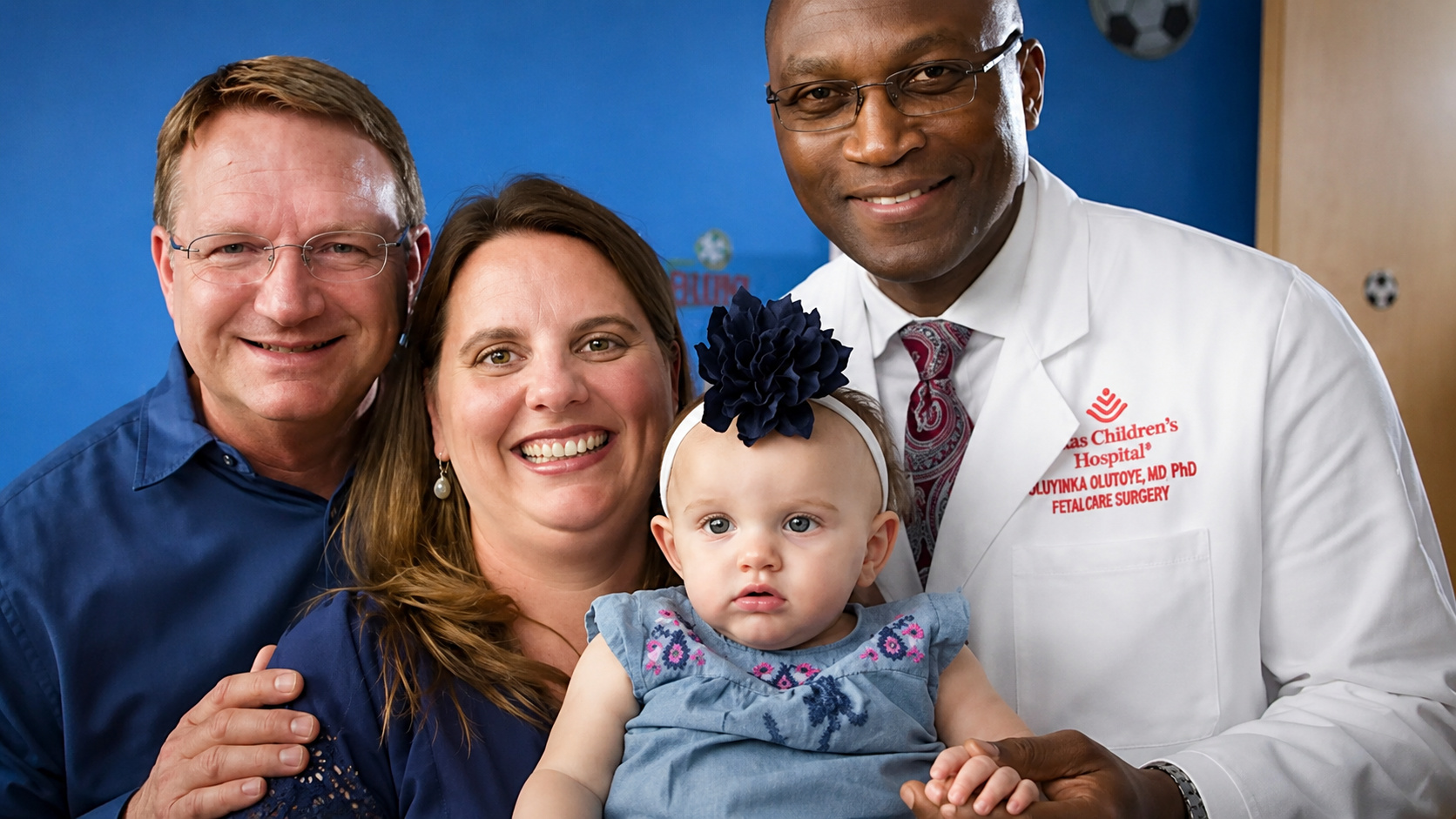
The case of Lynlee Boemer reveals a rare fetal surgery, a high-risk pregnancy, and a recovery marked by constant medical monitoring
A rare medical story drew attention by showing how Lynlee Boemer needed to be operated on even before officially being born. The girl was diagnosed in the womb with a sacrococcygeal teratoma, a tumor located in the coccyx region, which was growing rapidly and competing for blood with the baby. The case began in October 2015, when Margaret Boemer discovered she was pregnant with twins. After a miscarriage, only one baby survived, and the family discovered weeks later that they were expecting a girl. The joy, however, changed during an ultrasound when the parents noticed a strange image and the doctors confirmed the presence of the tumor.
Early diagnosis revealed serious risk for Lynlee
The diagnosis brought immediate concern because the tumor could already be seen at just 16 weeks of gestation. According to the original report, this type of tumor is the most common found in babies, but it is still rare, occurring in one in every 35,000 pregnancies. Additionally, the condition appears more frequently in girls and has an unknown cause. The biggest problem, however, was the tumor’s progression, which competed for blood with Lynlee and increased the risk of heart failure. Given the severity, Margaret and her husband were informed about the risks and began seeking an alternative to try to save their daughter.

Fetal surgery emerged as the only possible chance
The possibility found by the family was a fetal surgery, a rare and complex procedure that would partially remove Lynlee from the womb to remove part of the tumor. After that, she would be placed back in the mother’s belly so the pregnancy could continue. At that time, only four hospitals in the United States performed this type of intervention, and one of them was the Texas Children’s Hospital, in the same state where the family lived. It was there that the couple met Nigerian doctor Oluyinka Olutoye, a specialist in fetal surgery, who evaluated the case and explained that without intervention, Lynlee would likely die. Even so, the procedure was risky, as the doctors would operate on both mother and baby simultaneously.
-
Cold War Secret Plane Lost Engine Mid-Flight, Forcing Six to Parachute in Darkness; Wreckage Remains in Death Valley Mountains 73 Years Later

-
Opened in 1980, Shopping Recife was Pernambuco’s first mall and helped transform Boa Viagem into a bustling and sought-after district in southern Recife.
-
Tired of Monthly Water Supply, South African Villagers in Limpopo Connect Mountain Spring to Taps, Bringing Water to 5,000 People with $700 Initiative

-
Rare Case of Neurocysticercosis in Spain: 60-Year-Old Man’s Suspected Brain Cancer Turns Out to Be Tapeworm Cysts, Avoiding Invasive Cancer Treatments


Race against time changed the team’s plans
At 20 weeks of gestation, the tumor measured about 8 centimeters, almost the same size as Lynlee herself. The doctors, however, needed to wait until the baby’s body was better prepared to withstand incisions and sutures. The wait became increasingly delicate because the tumor grew along with her. At 23 weeks, a new check-up showed a rapid worsening of the condition, and the surgery needed to be performed immediately. In the operating room, about 20 professionals participated in the procedure. After an incision in Margaret’s abdomen and the use of ultrasound to avoid the placenta, the doctors accessed the uterus and exposed only the lower part of the baby’s body.
Delicate operation required transfusion during the procedure
Lynlee was extremely small at that moment, with only 11 centimeters and 530 grams. Therefore, the doctors needed to use magnifying lenses to perform the surgery. During the procedure, part of the tumor was removed, but the operation had to be interrupted for a transfusion, necessary to keep the baby alive. In the end, the surgery was considered successful. Dr. Oluyinka Olutoye stated that participating in the procedure was an honor, but emphasized that in such cases, the mothers are the true heroines because they put their own bodies at risk for their children. Margaret then remained at rest for the remainder of the pregnancy.

Official birth occurred in June 2016
The initial goal was to carry the pregnancy to the 38th week, but plans had to change again. On June 6, 2016, Lynlee was born by cesarean section at 36 weeks and 5 days. For Margaret, hearing her daughter’s cry, holding her, and kissing her was an emotional moment after so many risks. Eight days after birth, the baby underwent another surgery to remove the remaining tumor. The fetal operation, therefore, did not end all the care, as the family would still need to maintain constant medical follow-up due to the risk of the tumor returning.
Recovery turned fear into hope
At 1 year and 8 months, according to Margaret, Lynlee was doing very well, active and playful. The mother reported that her daughter ran everywhere, didn’t like to stay still, loved music, and showed joy in her daily life. Despite the need for monitoring, the recovery showed a very different outcome from the fear experienced during pregnancy. The girl who needed to be operated on inside the womb came to symbolize a rare story of survival, medical care, and family strength.
The case that impressed fetal medicine
Lynlee’s journey gained attention precisely because it involved an intervention that many people don’t even know is possible. The baby was partially removed from the womb, had part of the tumor removed, and then returned to continue the pregnancy. The official birth occurred months later, followed by another surgery. Today, the image described by the mother sums up the emotional dimension of the case: “Lynlee wakes up smiling every morning.”
In the face of such a rare story, how can we not see this case as one of the most impressive accounts of fetal medicine?


