Portuguese
  Spanish
The TomUS is a breast ultrasound scanner created at USP, uses sound waves without radiation and warm water to form sectional images, but still undergoes clinical validation before any widespread use in hospitals and can strengthen national medical technology.
On November 26, 2025, researchers from USP presented the TomUS, a breast ultrasound scanner created in Brazil. The prototype uses sound waves instead of radiation, forms sectional images, and has entered the clinical validation phase at the Hospital das Clínicas of the Faculty of Medicine of Ribeirão Preto at USP.
The information was published by Jornal da USP, a communication vehicle of the University of São Paulo. The project was born in the Department of Physics of the Faculty of Philosophy, Sciences, and Letters of Ribeirão Preto and brings together knowledge, design, and image processing developed in the country.
The TomUS is not a device available for general use in the healthcare network. It is a research prototype, created to demonstrate a technique, and still needs to be compared to already used exams before becoming a service tool.
-
Brazil Develops Its First Locally-Made 5G Radio Unit, Achieves Over 1.5 Gbps in Lab Tests, Paving the Way for Interoperable Equipment

-
El Niño Intensifies: Over 90% Chance of Persisting Until 2027, Raising Ocean Temperatures and Increasing Severe Weather Risks in Southern Brazil, Warns Federal Report

-
NASA Prepares Trio of Small Satellites to Track Tropical Storms and Capture Rapid Rainfall Development from Above the Clouds
-
$7,999 Isaac 1 Robot Aims to Tackle Household Chaos: Folds Clothes, Makes Beds, and Organizes, but Raises Privacy and Cost Concerns with Camera Reliance and Remote Human Assistance


In practice, the advancement places Brazilian engineering in an area where the country depends on ready-made equipment purchased abroad. The research can pave the way for new local solutions, but it does not change patient care now.
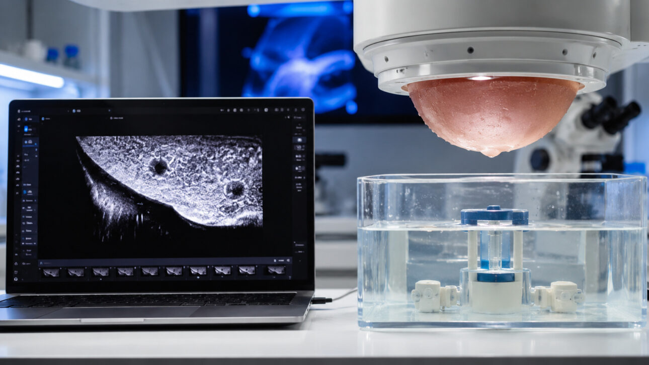
How the breast ultrasound scanner transforms waves into images
Ultrasound works with sound waves. They pass through the body, return to the device, and become information that helps build images of internal structures.
In the breast ultrasound scanner, these signals are gathered from many points. A computer program organizes the data and assembles a volume image, called a three-dimensional image, which can be viewed in sections.
Tomography is this: a way to observe tissue as if it could be divided into virtual slices. The doctor can look at each depth without needing to make cuts in the body.
Common ultrasound generates two-dimensional images and relies more on the movements of the person performing the exam. TomUS was built to automate the scanning and record breast tissue from various angles.
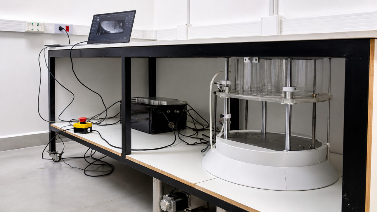
Warm water and robotic arm are part of the exam
In the exam, the patient lies face down on a table with an opening to position the breast. The area is immersed in warm water, which helps the sound waves to propagate through the tissue.
The system does not use breast compression. There is also no direct contact with the transducers, pieces that emit and receive the sound waves used to form the image.
A robotic arm performs a complete scan from multiple angles. The process is automated, which aims to reduce differences caused by the way each professional moves the device.
The information collection takes about five minutes. Then, a program transforms this data into images that show the interior of the tissue at different depths.
Slice images show the tissue from various angles
The images produced by TomUS allow the breast to be observed in slices. To better understand, think of a sliced loaf of bread: each part reveals a different depth without needing to move the rest.
This type of image can help the doctor navigate through the volume of the tissue. The assessment, however, still depends on clinical studies that show how the equipment behaves in real situations.
The system also allows the files to be gathered in a cloud database, so radiologists can analyze the images remotely. This possibility is part of the usage model studied by the researchers.
The important point is that the three-dimensional image does not, by itself, represent a diagnosis. It offers a new way to visualize the tissue, which still needs to be evaluated alongside already known methods.
Clinical validation defines the limits of TomUS before hospital use
The USP Journal, a communication vehicle of the University of São Paulo, detailed that TomUS has entered testing at the Hospital das Clínicas of the Ribeirão Preto Medical School. This stage includes a comparative analysis with traditional methods and a study of performance for diagnosis.
The initial tests involved physical models, which mimic body structures, and healthy volunteers. Now, the clinical phase needs to show how far the equipment can go and in which situations it can be used.
Jorge Elias Junior, a professor in the radiology department at the Ribeirão Preto Medical School, highlighted one of the questions that the tests need to answer: “We need to know, in clinical practice, what is the smallest lesion the equipment can detect.”
Lesions and tumors do not always appear the same way. The team still needs to verify, in real care, what the equipment’s limits are and in which situations it can be used.
Prototype does not replace mammography or common ultrasound
The TomUS does not automatically replace mammography, common ultrasound, or other already used exams. It is in the testing phase, and its possible application depends on the results of clinical validation.
The difference between a machine created for research and hospital-use equipment is significant. The prototype shows that the project works, but adoption in hospitals requires proof of performance, safety, and utility in medical routine.
The research opens a possibility, but the decision on broad use can only come after tests and comparisons with the methods already part of care.
National technology can reduce dependence on imported equipment
The research was developed by the Medical Innovation and Instrumentation Group and Ultrasound, linked to the Department of Physics of the Ribeirão Preto Faculty of Philosophy, Sciences, and Letters. The work includes the design of the device, image generation protocols, and data processing.
In November 2025, the ultrasound diagnostic equipment used in the country was imported. TomUS was built as a research alternative that keeps the necessary knowledge in Brazil to adapt and improve the system.
This does not mean that the cost of care will drop immediately. It also does not mean that hospitals will receive the device soon. The most direct impact, for now, is to expand Brazil’s capacity to create tools for the health area.
The development of a national medical device can facilitate the creation of new imaging protocols and other ultrasound-based applications. Each of these stages still requires its own tests before reaching people.
From the USP bench to the challenge of becoming a health equipment
The group responsible for TomUS is preparing to create a technology-based company in partnership with the Supera Innovation and Technology Park of Ribeirão Preto. The initiative brings together engineers, physicists, and computer professionals trained in the laboratory itself.
The goal is to continue improving the prototype and bring the research closer to the public and private health network. This path involves new clinical evaluations, technical development, and defining the best use for the system.
TomUS shows that the Brazilian university can build a complex solution that combines ultrasound, automation, and computer programs. The decisive part now is to transform this laboratory result into a safe, useful, and validated tool.
The importance of the research lies less in promising an immediate change and more in creating autonomy. When knowledge stays in the country, universities and professionals can develop improvements without relying solely on ready-made machines from abroad.
The breast ultrasound scanner created at USP already combines sound waves, warm water, automatic scanning, and cross-sectional images in a single prototype. The technology is still in clinical validation and does not replace the exams available to patients.
The next step is to prove, with clinical comparisons, when TomUS can be useful and what limits it will have. This process determines whether a laboratory research can responsibly reach hospitals.
What does Brazil need to do to safely bring research like TomUS from the laboratory to hospitals? Comment and share this post.